|

|
| Type |
Poster Presentation |
| Area |
Medicinal Chemistry |
| Room No. |
Grand Ballroom |
| Time |
10월 18일 (목요일) 11:00~12:30 |
| Code |
MEDI.P-297 |
| Subject |
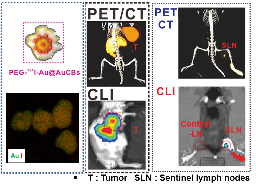
PEGylated crushed gold shell-radiolabeled core nanoballs for in vivo bio imaging application with dual positron emission tomography and cerenkov luminescent imaging |
| Authors |
Sangbong Lee, Jungwook Chin, Sung Jin Cho1,*
New Drug Development Center, Daegu Gyeongbuk Medical Innovation Foundation, Korea
1Daegu Gyeongbuk Medical Innovation Foundation, Korea
|
| Abstract |
|
Radioactive isotope labeled gold nanomaterials have potential biomedical imaging applications. Here, we report the synthesis and characterization of PEGylated crushed gold shell-radioactive iodide-124 labeled gold core nanoballs (PEG-124I-Au@AuCBs) for in vivo biomedical imaging application through combined positron emission tomography and cerenkov luminescent imaging (PET/CLI). PEG-124I-Au@AuCBs showed high stability and sensitivity in various pH solutions, serum, and in vivo conditions and were not toxic to tested cells. Combined PET/CLI clearly revealed disease lesions at 1 to 24 h after injection of particles, consistent with the biodistribution results. Taken together, the data provided strong evidence for the application of PEG-124I-Au@AuCBs as promising imaging agents in nuclear medicine imaging of various biological systems, particularly in diagnostics of various disease. |
|
|
| E-mail |
sangbongyi1@dgmif.re.kr |
|
;)